How an X-Ray Machine Works
No camera, no lens, no film in the modern ones. A wire boiled white-hot, electrons thrown across a vacuum at half the speed of light, and a spinning slab of tungsten that turns 99 per cent of it straight into heat — all to make the one per cent that casts a shadow through you.
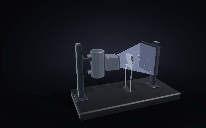
How does an X-ray machine work?
An X-ray machine boils electrons off a white-hot tungsten wire, hurls them across a vacuum at over half the speed of light into a spinning tungsten target, and lets the one percent of the impact that becomes radiation cast a shadow through whatever stands in its path. The image is a shadow, not a photograph.
Step 01 of 09
There is no camera in here
An X-ray machine takes no photograph. There is no lens anywhere in it and nothing in it responds to light. All it does is put a source of something that goes through solid objects at one end of a room, a detector at the other, and let you stand in between. Every X-ray image you have ever seen is a shadow — and this entire machine exists to make one small, hard, precisely-aimed place for that shadow to be cast from.
Step 02 of 09
A glass tube in a bath of oil
Take the housing off and there is startlingly little inside: one sealed glass envelope, about the size of a jam jar, floating in mineral oil. The oil has two jobs — carry the heat away, and hold a hundred thousand volts off the steel around it. That steel is lined with lead, because this tube sprays X-rays in every direction and only one of them is wanted. And inside the glass is a vacuum, pumped down hard enough that an electron can cross the whole tube without meeting a single atom.
Step 03 of 09
It starts by boiling a wire
At the top of the tube is the cathode, and it is nothing but a coil of tungsten wire roughly as thick as a human hair. Push a current through it and it sits at about 2,000 °C — white-hot, the way an old light bulb is — and at that temperature electrons stop being held by the metal and simply evaporate off it, hanging around the wire in a cloud. The little cup they sit in is charged negative, and since electrons are repelled by anything negative, the cup squeezes that cloud instead of letting it spread. How hard you heat the wire sets how many electrons you get: that is the milliamps on the control panel.
Step 04 of 09
Then drop them off a 100,000-volt cliff
Nothing has actually moved yet. Then the generator puts up to 120,000 volts between that wire and the metal target below it, and the cloud is gone — electrons crossing the gap at over half the speed of light. There is no air in the way to slow them or scatter them, so they arrive together, on a footprint about the size of a grain of rice. The kilovolts do not change how many electrons make the trip, only how hard each one lands, and that is what decides whether the X-rays coming out are soft enough to stop in skin or hard enough to cross a chest.
Step 05 of 09
Ninety-nine per cent of this is a heater
The electrons hit tungsten and stop dead. A few swerve past a nucleus instead of hitting one, and the sharper the swerve the more energy they shed as a single photon — braking radiation, which is most of the X-rays this machine makes. It is a dreadful way to make them. About one per cent of the energy leaves as X-rays; the other ninety-nine turns into heat, all of it landing on a patch you could cover with a full stop. Solid metal would melt in seconds. So the target is a disc, and the disc spins at three thousand revolutions a minute or more, dragging cold metal under the beam and smearing the damage around a whole ring instead of a point. The motor that turns it has no wires inside the vacuum: the copper rotor is sealed in the glass, and the stator coils outside drag it round through the wall.
Step 06 of 09
Only one way out
X-rays leave that spot in every direction at once, which is why the housing is lined with lead — anything not aimed at the port is stopped within a couple of millimetres of metal. What is left escapes through a window of beryllium, a metal so light that X-rays barely notice it is there. Then the collimator crops what remains: two pairs of lead blades slide in and out to cut the cone down to a rectangle exactly the size of the thing being imaged and not one centimetre bigger. That is not tidiness — every photon that lands outside the picture is dose spent for nothing. The mirror sitting at forty-five degrees in the beam folds a lamp along the same path, which is the only way to see where an invisible beam is pointing.
Step 07 of 09
The picture is a shadow
Now the beam crosses the room and meets something. Each step of this aluminium wedge is thicker than the last, and each one takes a bigger bite out of the photons crossing it — you can watch them stop arriving. What makes X-ray images legible is that absorption is brutally sensitive to what a material is made of: it climbs roughly with the cube of atomic number. That is the whole reason bone shows up. Calcium is atom-for-atom far heavier than the hydrogen, carbon and oxygen in everything packed around it, so it swallows photons its neighbours let straight through. A rib is a thicker, denser step.
Step 08 of 09
How a shadow becomes a file
The panel it lands on has no film in it and never did. First comes a grid of lead strips standing on edge, each one aimed back at the tube: a photon that came straight through passes between them, and a photon that bounced off something on the way — arriving at the wrong angle, carrying no useful information, only fog — hits a strip and dies there. Behind that is a layer of caesium iodide, grown in fine needles so the light cannot wander sideways, which flashes visibly every time an X-ray is absorbed in it. And behind that, a sheet of silicon photodiodes with a transistor at every pixel, reading out how much light each one caught. X-ray to light to charge to number, in about a second.
Step 09 of 09
Run it
Spin the anode up, heat the filament, open the blades, put a hundred thousand volts across the gap — and it is over in a few hundredths of a second. That is the strangest thing about this machine: it spends its whole life idling and cooling, and the exposure itself is shorter than a blink. A wire is boiled, electrons are thrown, a slab of tungsten gets briefly and violently hot, and one per cent of all that effort walks through you and leaves its shadow on the far wall.
The parts
- Cathode (filament) — A coil of tungsten wire about as thick as a human hair, heated to roughly 2,000 °C until electrons simply evaporate off it into the surrounding vacuum.
- Focusing cup — A negatively charged cup around the filament that squeezes the cloud of freed electrons into a tight stream instead of letting it drift apart.
- Anode — The spinning tungsten target the electrons slam into. It turns at 3,000+ RPM specifically so the impact point keeps changing — a stationary target would melt.
- Stator — The motor coils sealed outside the glass tube that spin the anode by induction, with no wire ever crossing the vacuum to reach it.
- Collimator — Two pairs of lead blades that crop the X-ray cone down to exactly the rectangle being imaged — every photon outside that rectangle is dose spent for nothing.
- Beryllium window — The one point in the lead-lined housing thin and light enough for X-rays to actually pass through, letting the beam out where it is wanted.
- Anti-scatter grid — A grid of lead strips standing on edge, aimed back at the tube, that catches photons arriving at the wrong angle — scattered light that would otherwise just fog the image.
- Scintillator — A layer of caesium iodide behind the grid that flashes visible light every time it absorbs an X-ray, handing the signal off to a silicon photodiode array underneath.
Numbers that matter
| Figure | Value | Note |
|---|---|---|
| Accelerating voltage | up to 120,000 V | the kilovolt drop electrons cross to reach the target |
| Electron speed at impact | over 50% of light speed | reached in a gap smaller than a coin |
| Filament temperature | ~2,000 °C | white-hot — hot enough to boil electrons directly off the metal |
| Anode rotation speed | 3,000+ RPM | spreads the heat around a ring instead of melting a single point |
| Energy → X-ray conversion | ~1% | the other ~99% becomes heat, which is why the anode has to spin at all |
| Typical exposure duration | hundredths of a second | the machine spends nearly all its life idling and cooling |
| Dose, one chest X-ray | ~0.1 mSv | about 10 days of ordinary background radiation |
Common questions
Is it safe to get an X-ray?
A single chest X-ray delivers around 0.1 millisievert — about ten days’ worth of the background radiation everyone absorbs anyway just from cosmic rays and the ground underfoot. It’s not zero, which is why exposures are only ordered when there’s a real medical reason, but the dose itself is small next to what a body handles from the environment every year regardless.
Does the lead apron protect your whole body?
No — the apron most rooms use is sleeveless and stops above the knees, leaving the arms, shoulders and head uncovered. It’s become enough of a coverage myth that the American Association of Physicists in Medicine no longer even recommends shielding reproductive organs during most imaging: the dose reduction is marginal, and studies have found the shields themselves misplaced roughly half the time, sometimes covering the very bone that was supposed to be imaged.
Why do bones show up so clearly but soft tissue barely does?
Because how much a material absorbs climbs roughly with the cube of its atomic number. Calcium is atom-for-atom far heavier than the hydrogen, carbon and oxygen that make up most soft tissue, so bone swallows photons that everything around it lets straight through — that contrast is the entire image.
Why does the target spin instead of just sitting still?
Because ninety-nine percent of the electron beam’s energy becomes heat, not X-rays, landing on a patch of metal smaller than a fingertip. A stationary target would melt in seconds; spinning it at 3,000+ RPM drags cold metal continuously under the beam and spreads that heat around a whole ring instead.
Can X-rays pass through anything?
No — that’s exactly why the tube housing is lined with lead and why the room itself is often shielded. Dense, high-atomic-number materials like lead absorb X-rays almost completely, which is the same property that makes bone visible against soft tissue, just taken to an extreme.
What goes wrong
What actually breaks inside an X-ray tube?
Mostly thermal fatigue. Every exposure heats the tungsten target and lets it cool again, and that repeated cycling eventually opens micro-cracks in the target surface and the stem behind it. Rotor bearing wear compounds it — an imbalanced disc vibrates, and vibration accelerates exactly the stem-cracking that thermal cycling already causes.
Why do older X-ray tubes start arcing?
Over years of operation, a thin film of vaporized tungsten gradually deposits on the inside of the glass envelope. Once that film gets thick enough, it forms a conductive path across the insulator, and the tube starts arcing internally — a slow-motion failure that a tube can accumulate toward for years before it becomes noticeable.
Can a protective shield make an exposure worse?
Occasionally, yes. When a shield is placed even slightly wrong, it can obscure the exact anatomy the X-ray was ordered to check — and a study found that happening in roughly half of pelvic exposures. The result isn’t a safer image, it’s a repeat exposure, which is part of why shielding guidance has shifted away from routinely using it at all.